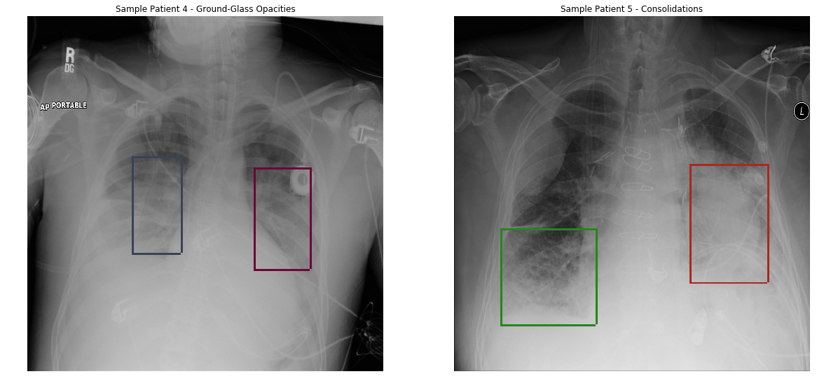
Pnumonia Chest Xray

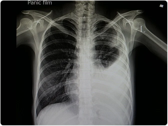
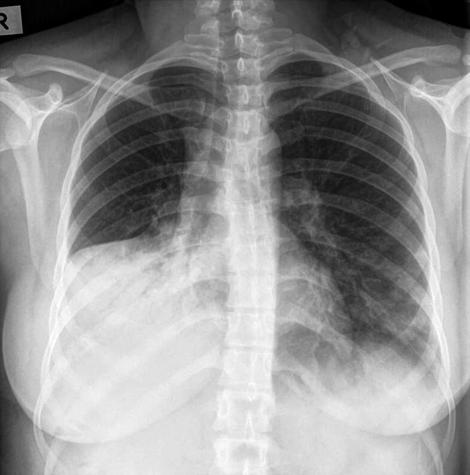
Pneumonia may have an associated parapneumonic effusion.
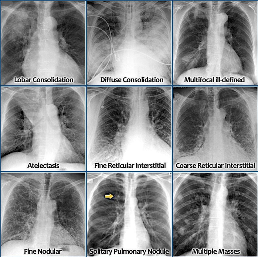
Pnumonia chest xray. Lobular often staphlococcus multifocal patchy sometimes without air bronchograms. Pneumonia is a general term in widespread use defined as infection within the lung. Terminology pneumonia is in contrast to pneumonitis which is inflammation of the pulmonary inter. Lobar classically pneumococcal pneumonia entire lobe consolidated and air bronchograms common.
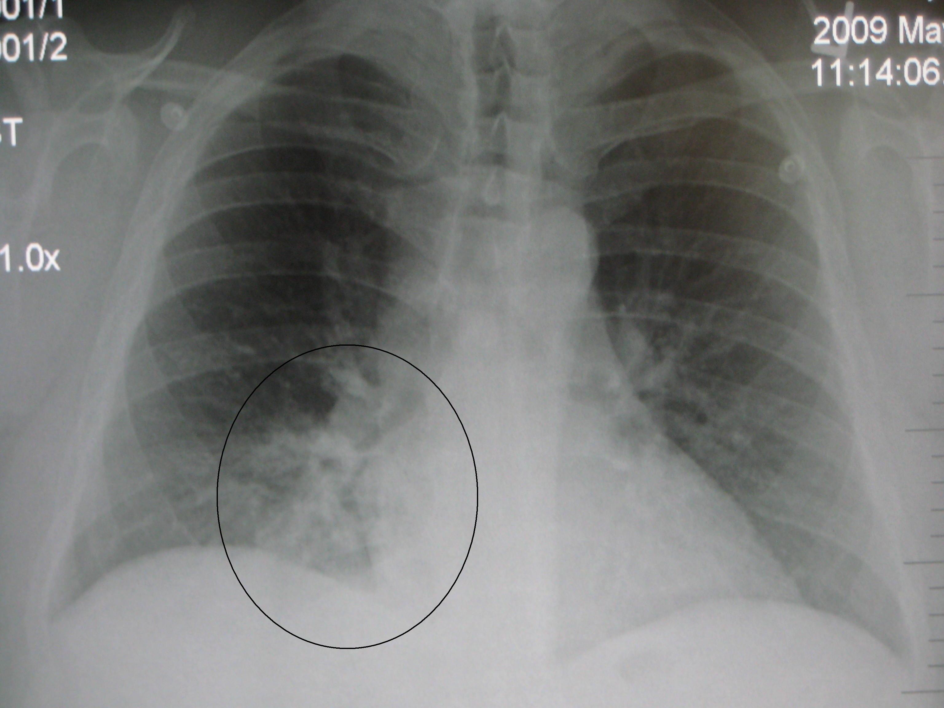
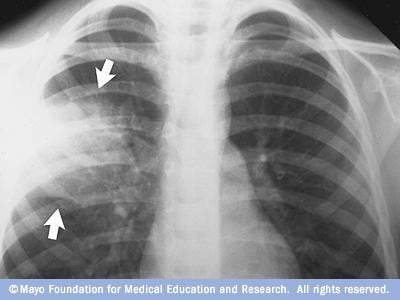
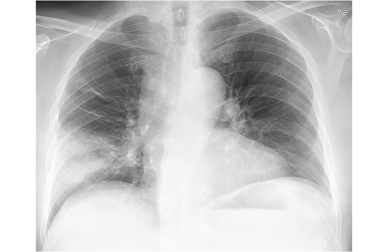
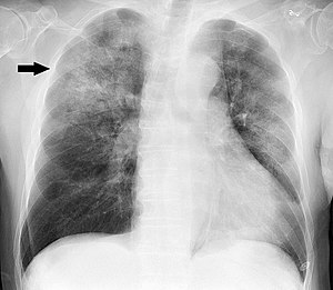
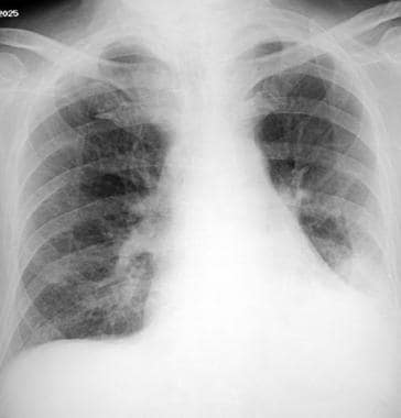
This chest x ray shows an area of lung inflammation indicating the presence of pneumonia. You can see three columns. It is due to material usually purulent filling the alveoli. Streptococcus pneumoniae is by far the most common causative organism.
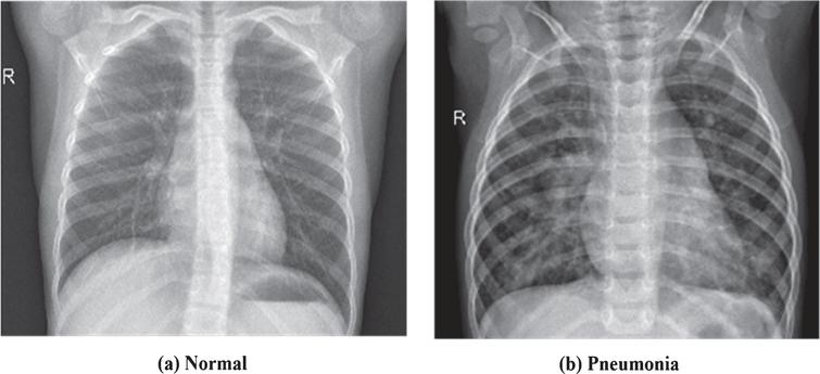
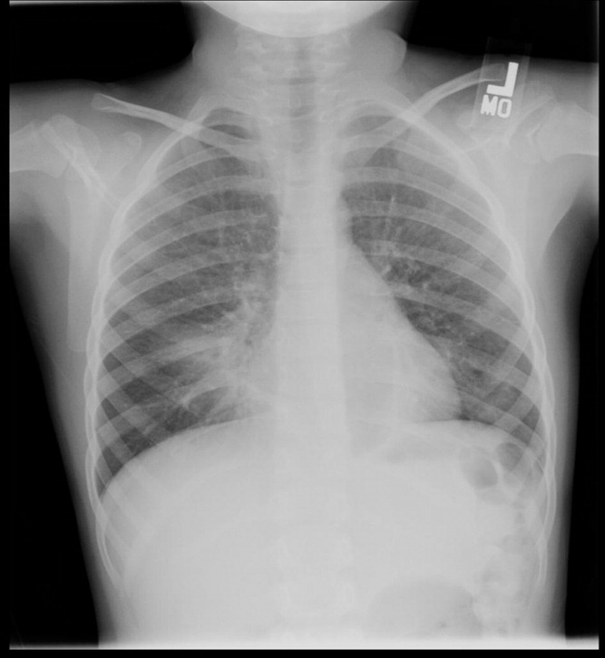
Each row in the table represents one image of a chest x ray. Chest x ray images anterior posterior were selected from retrospective cohorts of pediatric patients of one to five years old from guangzhou women and children s medical center guangzhou. Pneumonia is characterised by exudation and consolidation into the alveoli and in the u k. Class is either 0 or a 1 0 meaning that the lung is healthy and 1 meaning that the lung has pneumonia.
There are 5 863 x ray images jpeg and 2 categories pneumonia normal. All chest x ray imaging was performed as part of patients routine clinical care. As such coders need to watch for it in documentation. The index is a label for the image that tells us which image in the dataset we are looking at.
Class split and index. Your doctor will start by asking about your medical history and doing a physical exam including listening to your lungs with a stethoscope to check for abnormal bubbling or crackling sounds that suggest pneumonia.









:max_bytes(150000):strip_icc()/covid-19-pneumonia-12-20adbdbe7ee54f7784689c3b1ede2d1c.jpg)
