Pnuemothorax Xray

Pneumothorax Radiology Case Radiopaedia Org

Chest Radiology

Pneumothorax Radiology Case Radiopaedia Org

How To Identify Pneumothorax On A Chest X Ray Youtube

Pneumothorax Pulmonary Disorders Merck Manuals Professional Edition

Chest Xray In Pneumothorax
When a pneumothorax is suspected in a supine patient confirmation can be readily obtained with a lateral decubitus view.
Pnuemothorax xray. This air pushes on the outside of your lung and makes it collapse. See pneumothorax in a supine patient and pneumothorax is one cause of a transradiant hemithorax. Regression analysis based on volume measurements from helical ct. Take our chest x ray essentials course and learn how to interpret chest x rays like a pro.
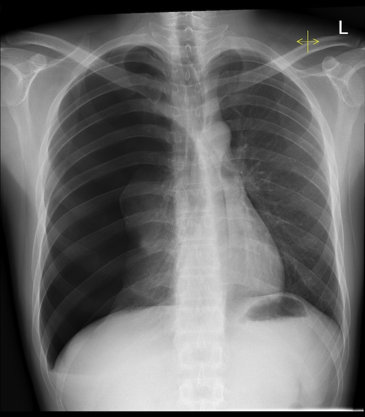
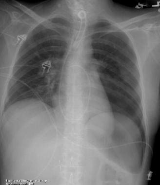
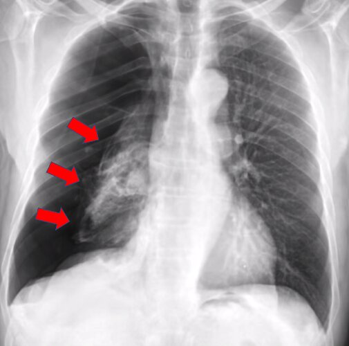
A pneumothorax occurs when air leaks into the space between your lung and chest wall. Pneumothorax can be a complete lung collapse or a collapse of only a portion of the lung. The example shown is a complete left pneumothorax. In this video you ll learn to identify when radiological pleura is abnormal and the key signs to look out for when trying to diagnose a pneumothorax.
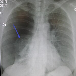
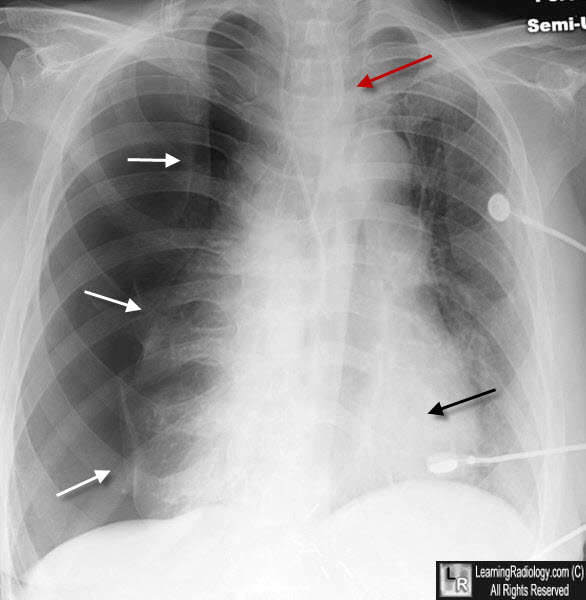
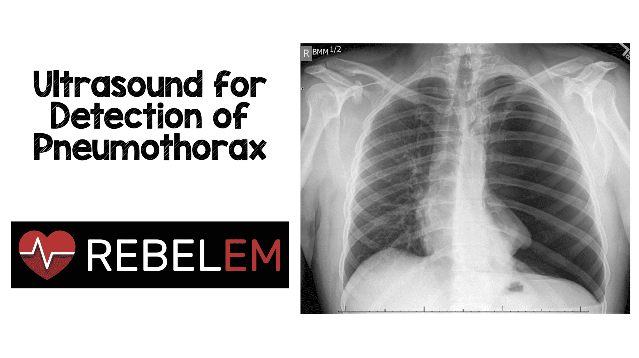
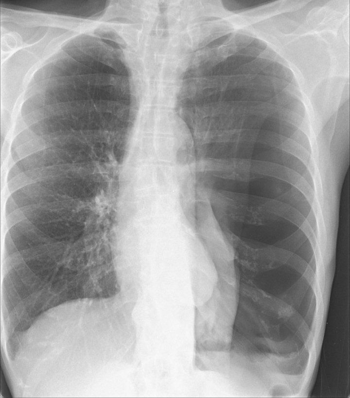
The lungs can be seen to reach the inner edge of the thoracic wall arrows. Ajr am j roentgenol 1995. Use this chest x ray as a normal reference for this gallery. Ultrasound imaging also may be used to identify a pneumothorax.
Ultrasound m mode can be used to determine movement of the lung within the rib interspace. Quantification of pneumothorax size on chest radiographs using interpleural distances. Collins cd lopez a mathie a et al. Ct may also be useful and has been shown to identify 50 to 64 of occult pneumothoraces in the trauma setting.
A pneumothorax is generally diagnosed using a chest x ray. In some cases a computerized tomography ct scan may be needed to provide more detailed images. The two lungs meet in the middle at the posterior junctional line arrowheads and the anterior junctional line not clearly visible. A pneumothorax noo moe thor aks is a collapsed lung.
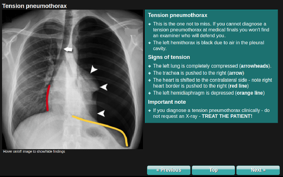
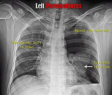
It is a life threatening occurrence requiring both rapid recognition and prompt treatment to avoid a cardiorespiratory arrest. These are white branching structures on the x ray if there are not vessels there may be a pneumothorax if both these findings occur measure how deep the pneumothorax is and check whether there is any mediastinal shift. How to identify pneumothorax on a chest x ray. Chest x ray showing the features of pneumothorax on the left side of the person right in image a plain chest radiograph ideally with the x ray beams being projected from the back posteroanterior or pa and during maximal inspiration holding one s breath is the most appropriate first investigation.
Want to master chest x ray interpretation. Chest radiograph a poor method for determining the size of a pneumothorax. Tension pneumothoraces occur when intrapleural air accumulates progressively in such a way as to exert positive pressure on mediastinal and intrathoracic structures.
Tension Pneumothorax Radiology At St Vincent S University Hospital
Pneumothorax Wikipedia

Pneumothorax Radiology In Two Minutes Youtube

Chest Xray In Pneumothorax

Pneumothorax X Ray Stock Image M240 0471 Science Photo Library

Chest X Ray Showing Large Right Pneumothorax With Collapsed Lung Download Scientific Diagram

Chest X Ray Pneumothorax Detection Curacloud

Pneumothorax Radiology Reference Article Radiopaedia Org

Chest Radiology
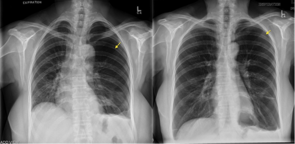
Pneumothorax Expiration Radiology At St Vincent S University Hospital

Pneumothorax Undergraduate Diagnostic Imaging Fundamentals
Pneumothorax Imaging Practice Essentials Radiography Computed Tomography

Pin On X Ray
Pneumothorax Wikipedia
Siim Acr Pneumothorax Segmentation Kaggle

Tension Pneumothorax Radiology Key
Learningradiology Tension Pneumothorax Ptx Spontaneous Shift Imaging

A Plain Chest X Ray Film Shows Massive Right Pneumothorax With Download Scientific Diagram
3
Ultrasound For Detection Of Pneumothorax Rebel Em Emergency Medicine Blog

Re Expansion Pulmonary Oedema In Pneumothorax Bmj Case Reports
Artificial Intelligence That Reads Chest X Rays Is Approved By Fda Uc San Francisco
Ai System More Accurately Identifies Collapsed Lungs Using Chest X Rays
