Pnuemothorax Chest X Ray

Chest Radiology

Chest Xray In Pneumothorax

How To Identify Pneumothorax On A Chest X Ray Youtube

Pneumothorax Radiology Case Radiopaedia Org
Apical Pneumothorax Radiology Case Radiopaedia Org

Chest Xray In Pneumothorax
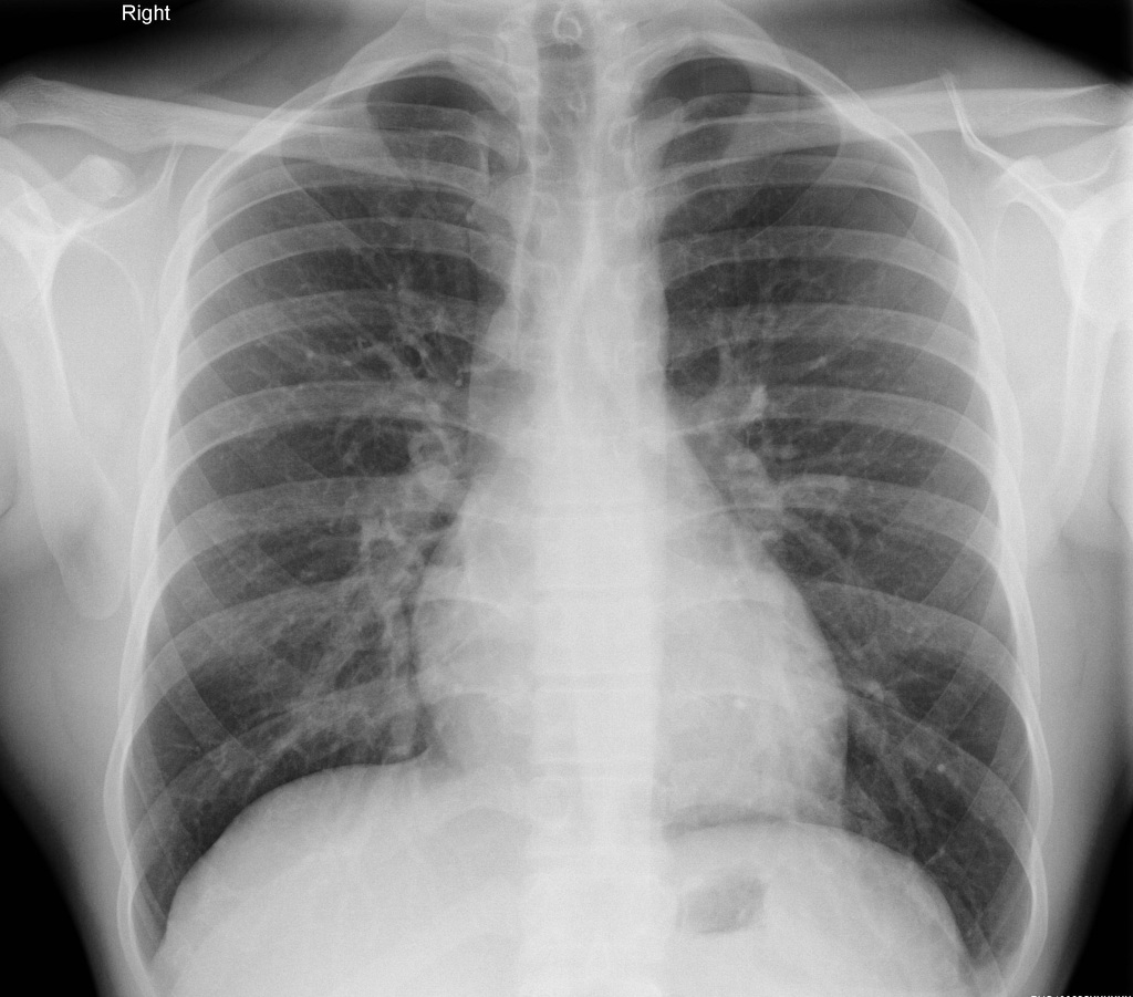
The lungs can be seen to reach the inner edge of the thoracic wall arrows.
Pnuemothorax chest x ray. The two lungs meet in the middle at the posterior junctional line arrowheads and the anterior junctional line not clearly visible. Pneumothorax occurs when air leaks from inside of the lung to the space between the lung and the chest wall. Regression analysis based on volume measurements from helical ct. Related records engdahl o toft t boe j.
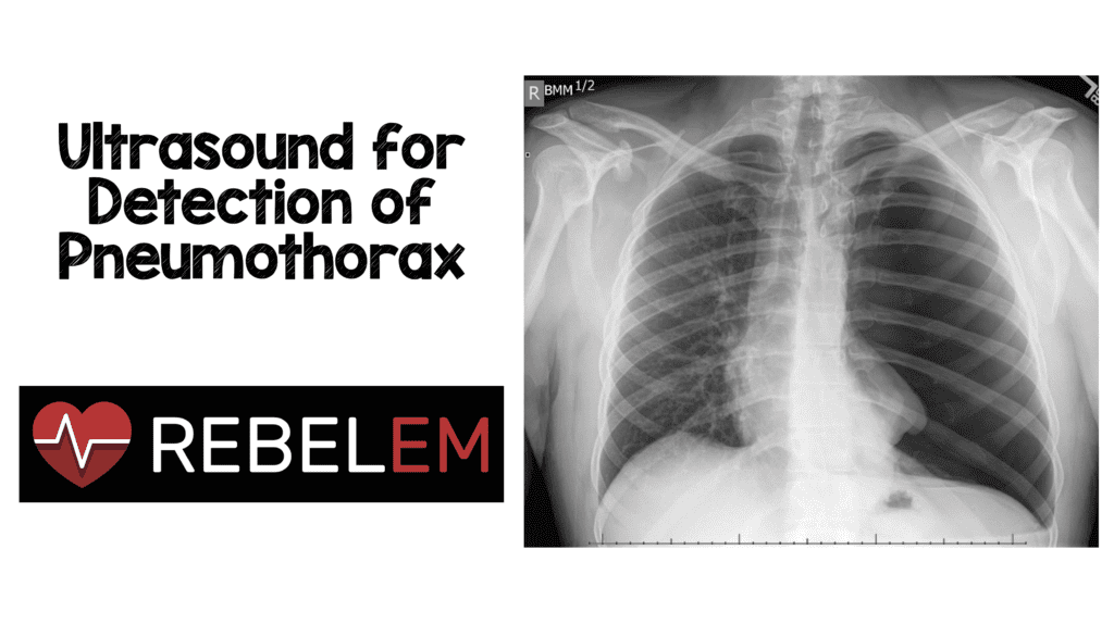
Pneumothoraces refers to the presence of gas air in the pleural space when this collection of gas is constantly enlarging with resulting compression of mediastinal structures it can be life threatening and is known as a tension pneumothorax if no tension is present it is a simple pneumothorax for those pneumothoraces occurring in neonates see the article on neonatal. Gold standard in pneumothorax indicated where chest xray cannot distinguish bleb in copd from pneumothorax in those with secondary spontaneous pneumothorax due to blebs contralateral blebs are seen in 50 of cases these contralateral blebs have a 25 chance of future secondary pneumothorax. Chest radiograph a poor method for determining the size of a pneumothorax. Ultrasound imaging also may be used to identify a pneumothorax.
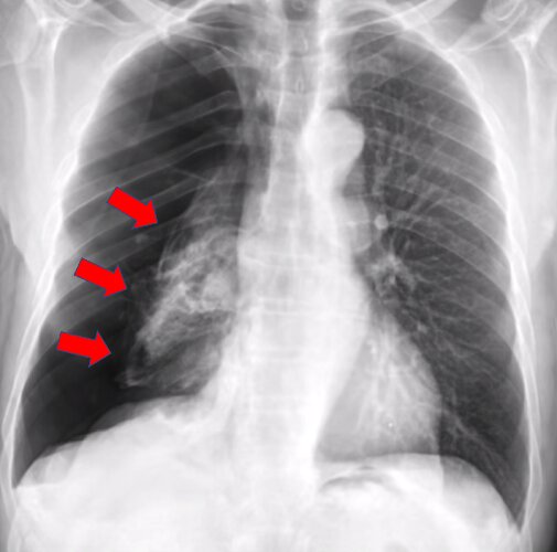
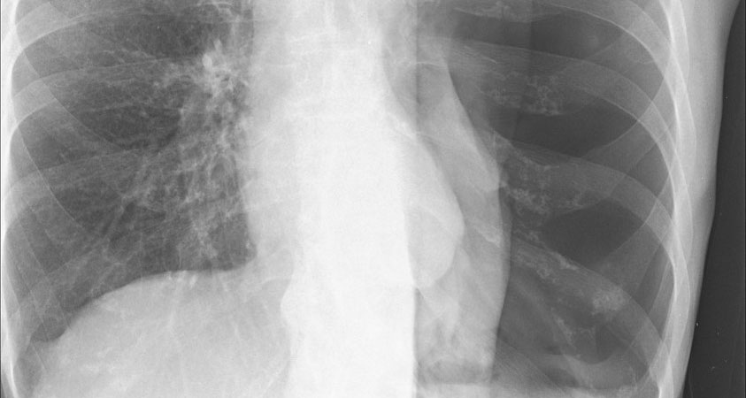
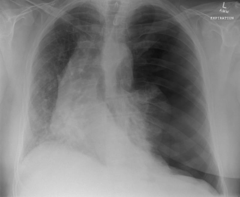
Was this page helpful. The dark side of the chest right side of the picture is filled with air that is outside of the lung tissue. Quantification of pneumothorax size on chest radiographs using interpleural distances. Zhang et al pooled 20 studies that compared ultrasound chest x ray or both against a reference standard usually ct scan for the diagnosis of pneumothorax.
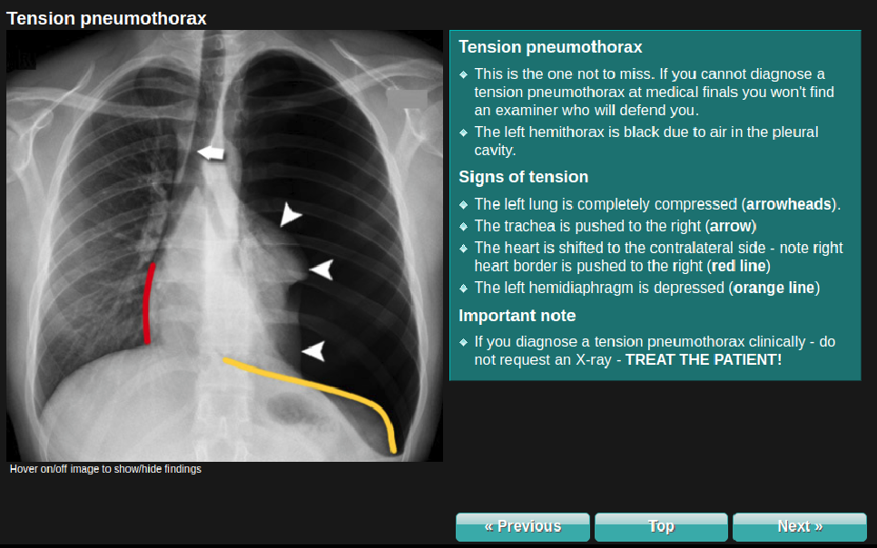
In some cases a computerized tomography ct scan may be needed to provide more detailed images. In this video you ll learn to identify when radiological pleura is abnormal and the key signs to look out for when trying to diagnose a pneumothorax. Ajr am j roentgenol 1995. A tension pneumothorax occurs due to the progressive accumulation of intrapleural gas in thoracic cavity caused by a valve effect during inspiration expiration.
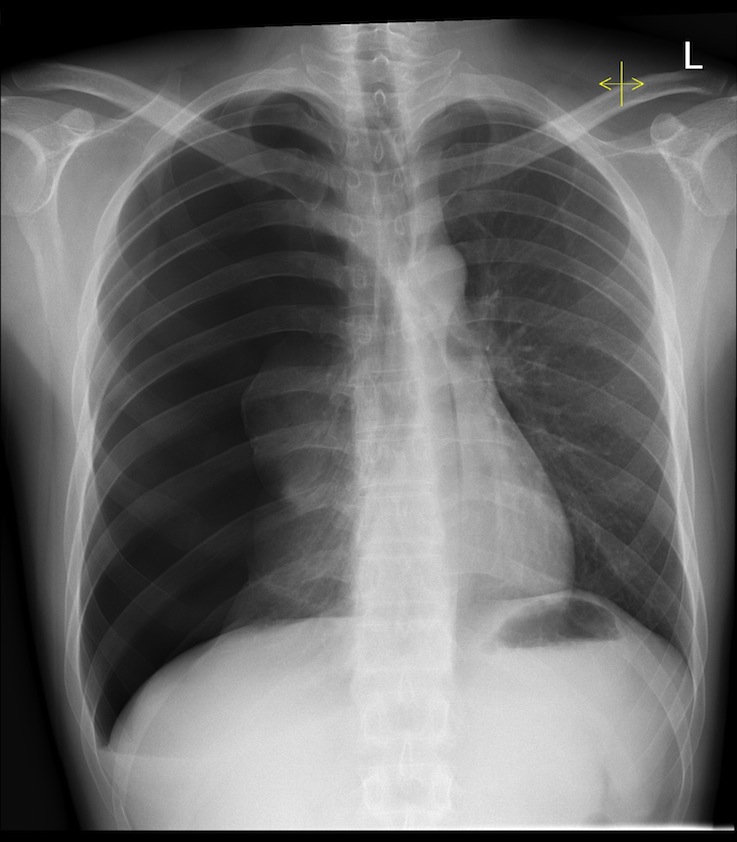
Collins cd lopez a mathie a et al. If you diagnose a tension pneumothorax clinically do not request an x ray treat the patient. Want to master chest x ray interpretation. Use this chest x ray as a normal reference for this gallery.
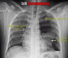
Chest x ray had a pooled sensitivity of 52 and specificity 99 for diagnosis of pneumothorax. Take our chest x ray essentials course and learn how to interpret chest x rays like a pro. In this situation the ipsilateral lung will if normal collapse completely although a less than normally compliant lung may remain partially inflated. How to identify pneumothorax on a chest x ray.
Ai System More Accurately Identifies Collapsed Lungs Using Chest X Rays

Pneumothorax Wikipedia

Pneumothorax Undergraduate Diagnostic Imaging Fundamentals

Chest X Ray Showing Large Right Pneumothorax With Collapsed Lung Download Scientific Diagram
Tension Pneumothorax Radiology At St Vincent S University Hospital

Pneumothorax Radiology Case Radiopaedia Org
Boring Question How Does The Sensitivity Specificity Of Lung Ultrasound Compare To Plain Films In Diagnosing Pneumothorax Canadiem

Chest X Ray Showing Bilateral Pneumothorax With A Flattened Diaphragm Download Scientific Diagram

Pneumothorax Pulmonary Disorders Merck Manuals Professional Edition

Chest X Ray Pneumothorax Or No Pneumothorax Youtube

Fda Clears Software To Spot Collapsed Lung In Chest X Rays Medgadget
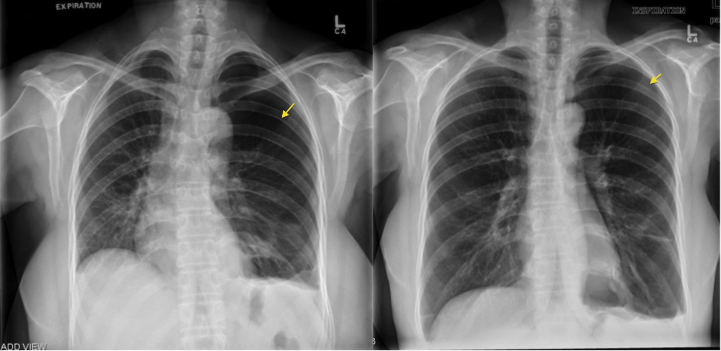
Pneumothorax Expiration Radiology At St Vincent S University Hospital
Artificial Intelligence That Reads Chest X Rays Is Approved By Fda Uc San Francisco
Pneumothorax Wikipedia

Pin On X Ray

Pneumothorax

Chest Radiology
Ultrasound For Detection Of Pneumothorax Rebel Em Emergency Medicine Blog
3

Chest X Ray Pneumothorax Detection Curacloud
Tension Pneumothorax An Alternative View Litfl
Siim Acr Pneumothorax Segmentation Kaggle

Pneumothorax Radiology Reference Article Radiopaedia Org
