Pnuemonia Cxr

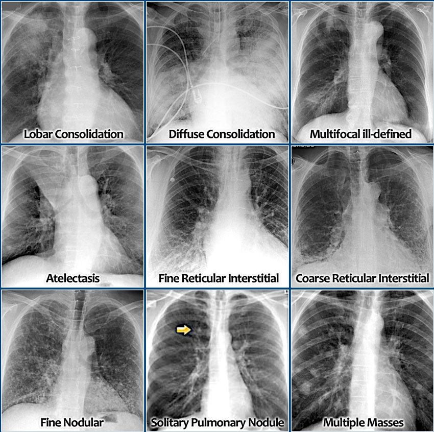
Consolidation seen in a non lobar distribution should raise the suspicion of atypical organisms.
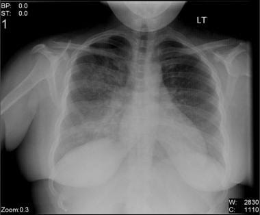
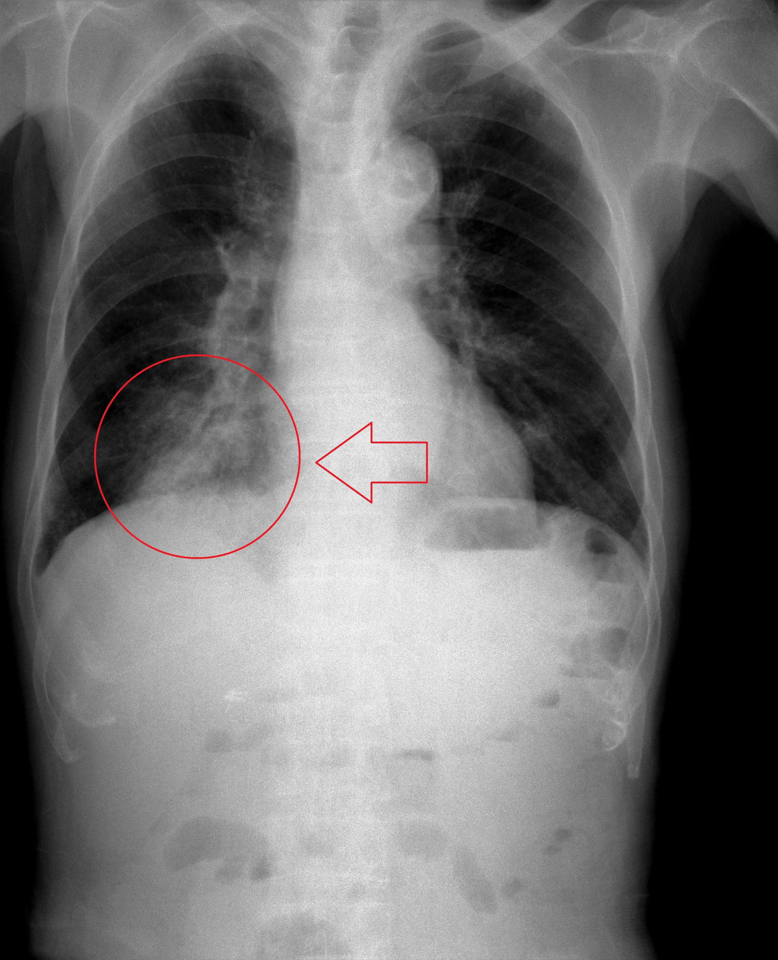
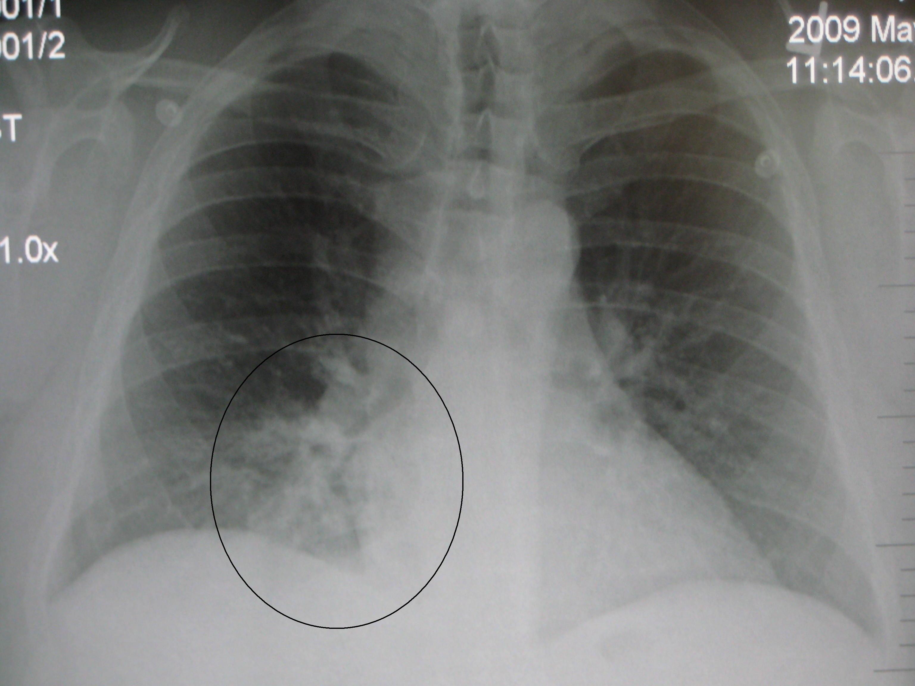
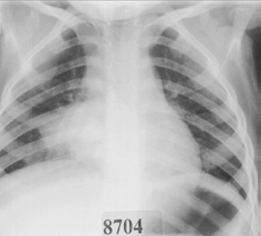
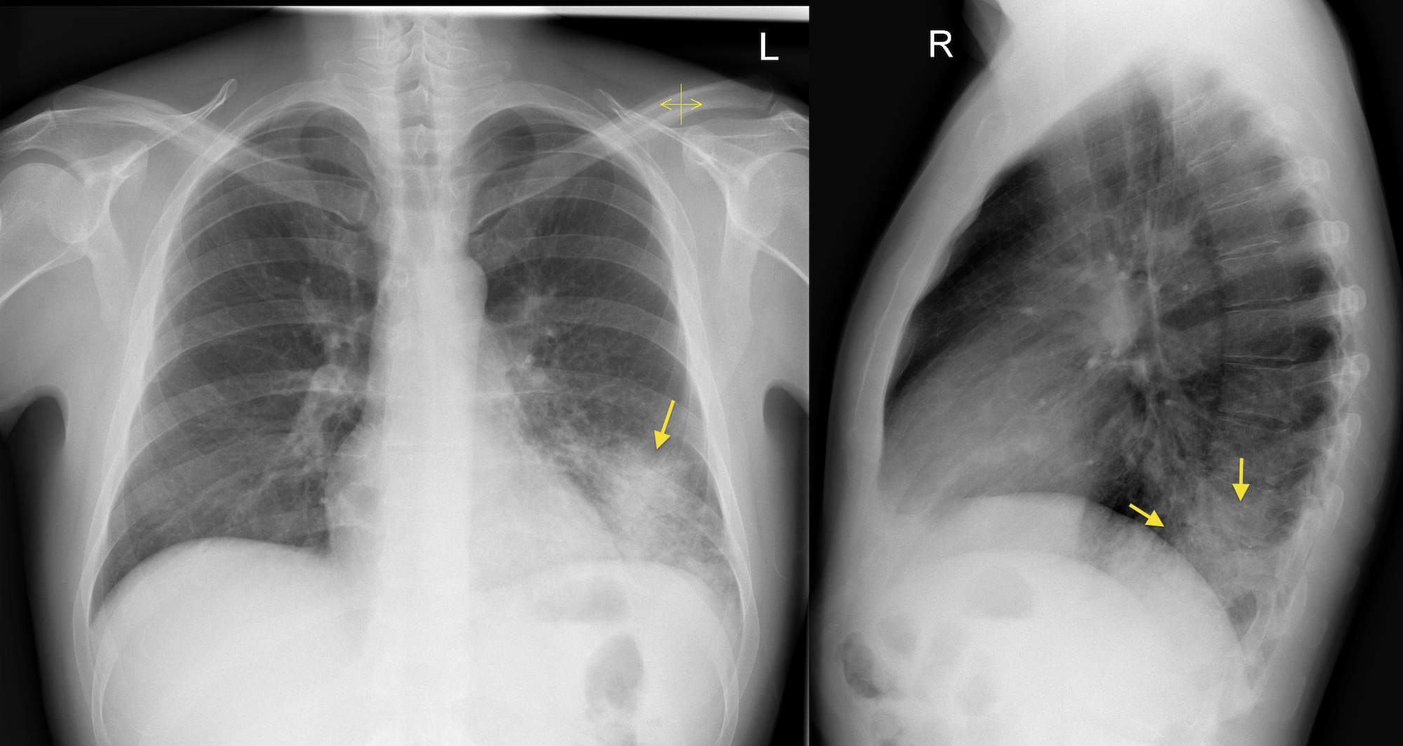
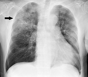
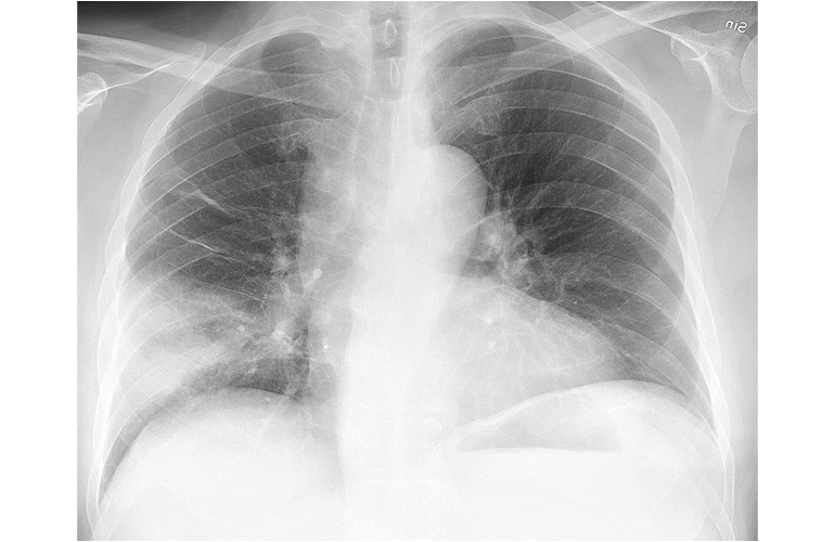
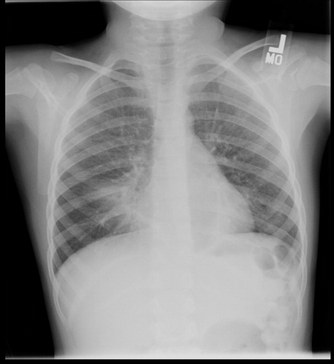
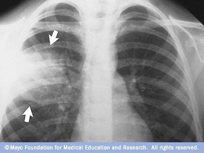
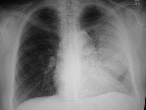
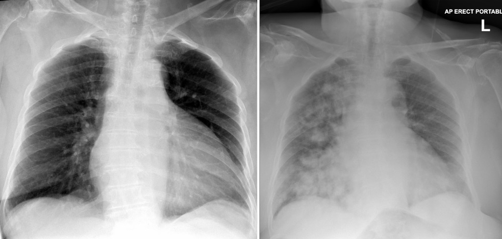
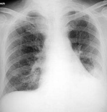
Pnuemonia cxr. If pneumonia is suspected your doctor may recommend the following tests. Your doctor may conduct a physical exam and use chest x ray chest ct chest ultrasound or needle biopsy of the lung to help diagnose your condition. Bronchopneumonia appears on a chest x ray as bilateral patchy shadows and typically predominates at the lung bases. Terminology pneumonia is in contrast to pneumonitis which is inflammation of the pulmonary inter.
Pneumonia is an infection that affects one or both lungs. Pneumonia is a serious complication of the new coronavirus also known as covid 19. This lung illness may cause severe breathing problems that put you in the hospital. Bacteria viruses or fungi may cause pneumonia.
Tap on off image to show hide findings. This patient with known hiv infection has subtle consolidation in the mid zones bilaterally. Pneumonia is an infection that causes inflammation in one or both of the lungs and may be caused by a virus bacteria fungi or other germs. Hover on off image to show hide findings.
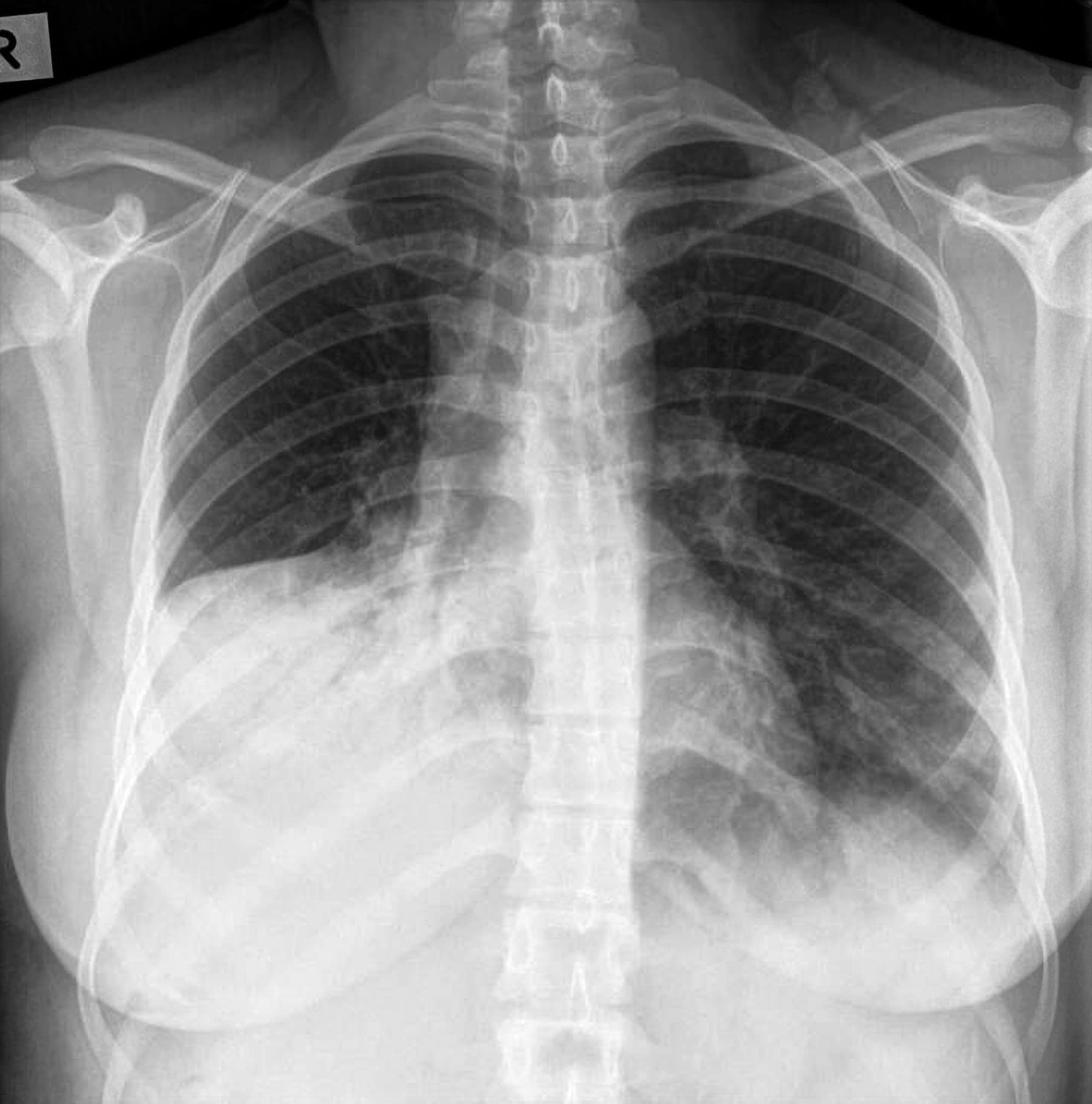
A variety of organisms including bacteria viruses and fungi can cause pneumonia. The air sacs may fill with fluid or pus purulent material causing cough with phlegm or pus fever chills and difficulty breathing. Symptoms can range from mild to serious and may include a cough with or without mucus a slimy substance fever chills and trouble breathing. Lobar pneumonias have specific appearances which can be explained by the anatomical relationships of the lobes of the lung to their surrounding structures.
It is due to material usually purulent filling the alveoli. Chest x ray showing pneumonia your doctor will start by asking about your medical history and doing a physical exam including listening to your lungs with a stethoscope to check for abnormal bubbling or crackling sounds that suggest pneumonia.








:max_bytes(150000):strip_icc()/covid-19-pneumonia-12-20adbdbe7ee54f7784689c3b1ede2d1c.jpg)
