Pneumonia Chest X Ray
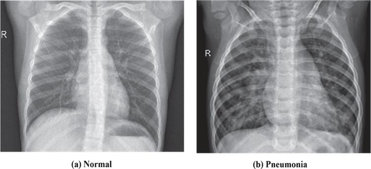
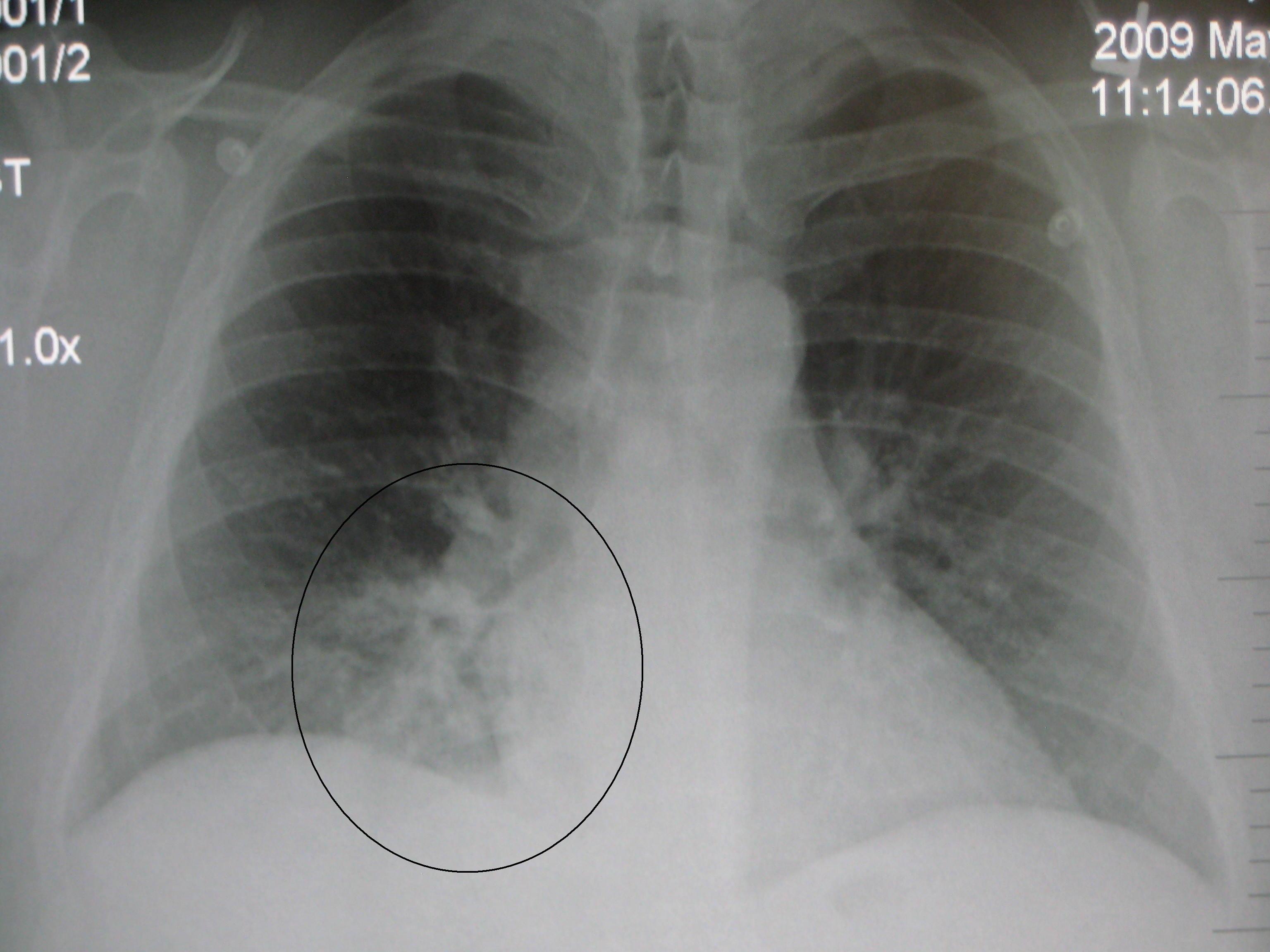
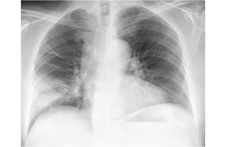
The larger image shows a left diaphragm silhouette sign indicating a left lower lobe bacterial pneumonia.
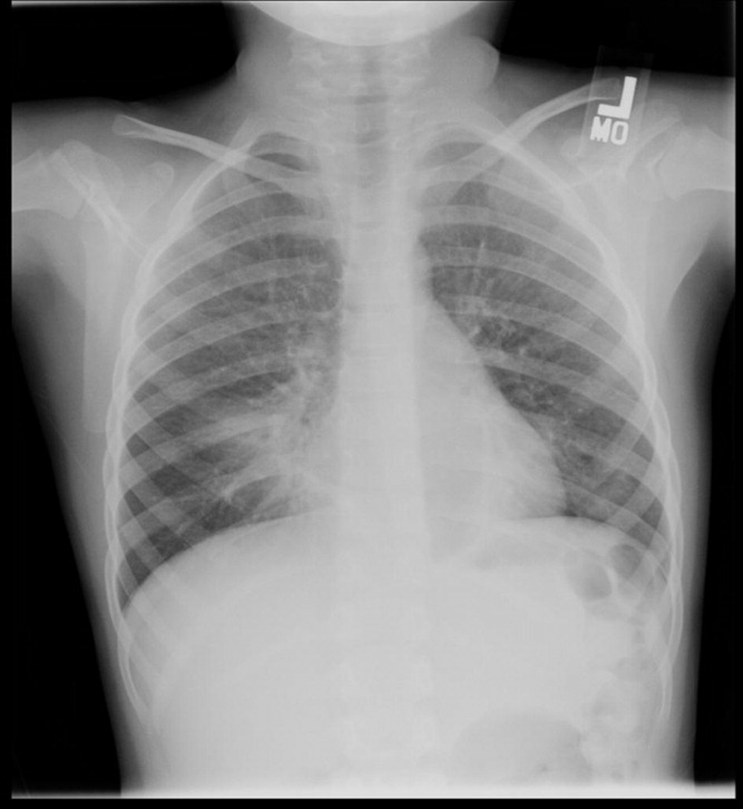
Pneumonia chest x ray. Chest x ray classification for pneumonia. Pneumonia is typically diagnosed based on a combination of physical signs and a chest x ray. Chest x rays are the initial modality of investigation in the majority of cases and a sound understanding of the chest x ray features of pneumonia is vital for all front line clinicians that encounter and treat it. In adults with normal vital signs and a normal lung examination the diagnosis is unlikely.
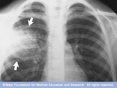
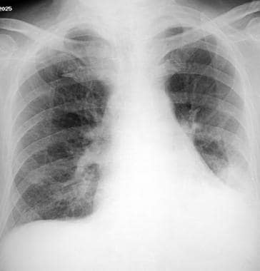
The type of pneumonia is sometimes characteristic on chest x ray. This chest x ray shows an area of lung inflammation indicating the presence of pneumonia. It is due to material usually purulent filling the alveoli. An x ray exam will allow your doctor to see your lungs heart and blood vessels to help determine if you.
Your doctor will start by asking about your medical history and doing a physical exam including listening to your lungs with a stethoscope to check for abnormal bubbling or crackling sounds that suggest pneumonia. To diagnose pneumonia your doctor will review your medical history perform a physical exam and order diagnostic tests such as a chest x ray. It has also resulted in reduced use of invasive procedures. A ct scan of the chest may be done to see finer details within the lungs and detect.
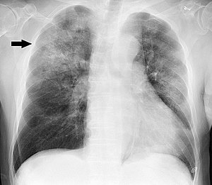
This information can help your doctor determine what type of pneumonia you have. Streptococcus pneumoniae is by far the most common causative organism. Your doctor could order a chest x ray for a variety of reasons including to assess injuries. The inset radiograph shows a right upper lobe pneumonia.
Chest x rays can also determine if you have fluid in your lungs or fluid or air surrounding your lungs. Ct of the lungs. However the underlying cause can be difficult to confirm as there is no definitive test able to distinguish between bacterial and non bacterial origin. Terminology pneumonia is in contrast to pneumonitis which is inflammation of the pulmonary inter.
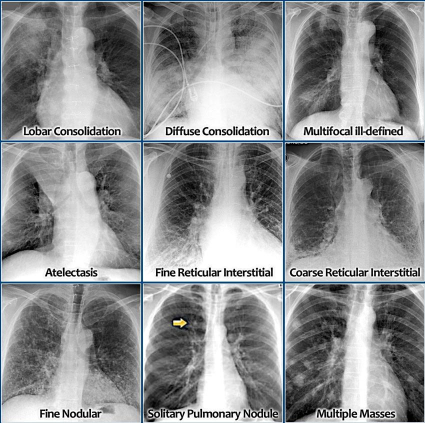
And with increase in data the burden in medical experts examining that data increases. One or more of the following tests may be ordered to evaluate for pneumonia. Lobar classically pneumococcal pneumonia entire lobe consolidated and air bronchograms common lobular often staphlococcus multifocal patchy sometimes without air bronchograms interstitial viral or mycoplasma. Pneumonia is a general term in widespread use defined as infection within the lung.
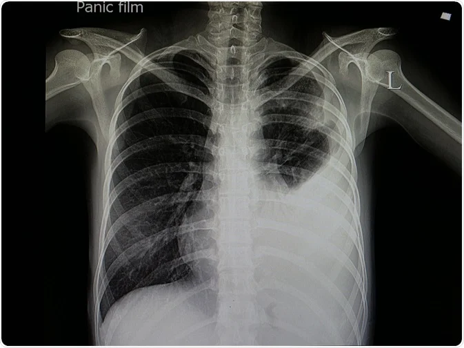
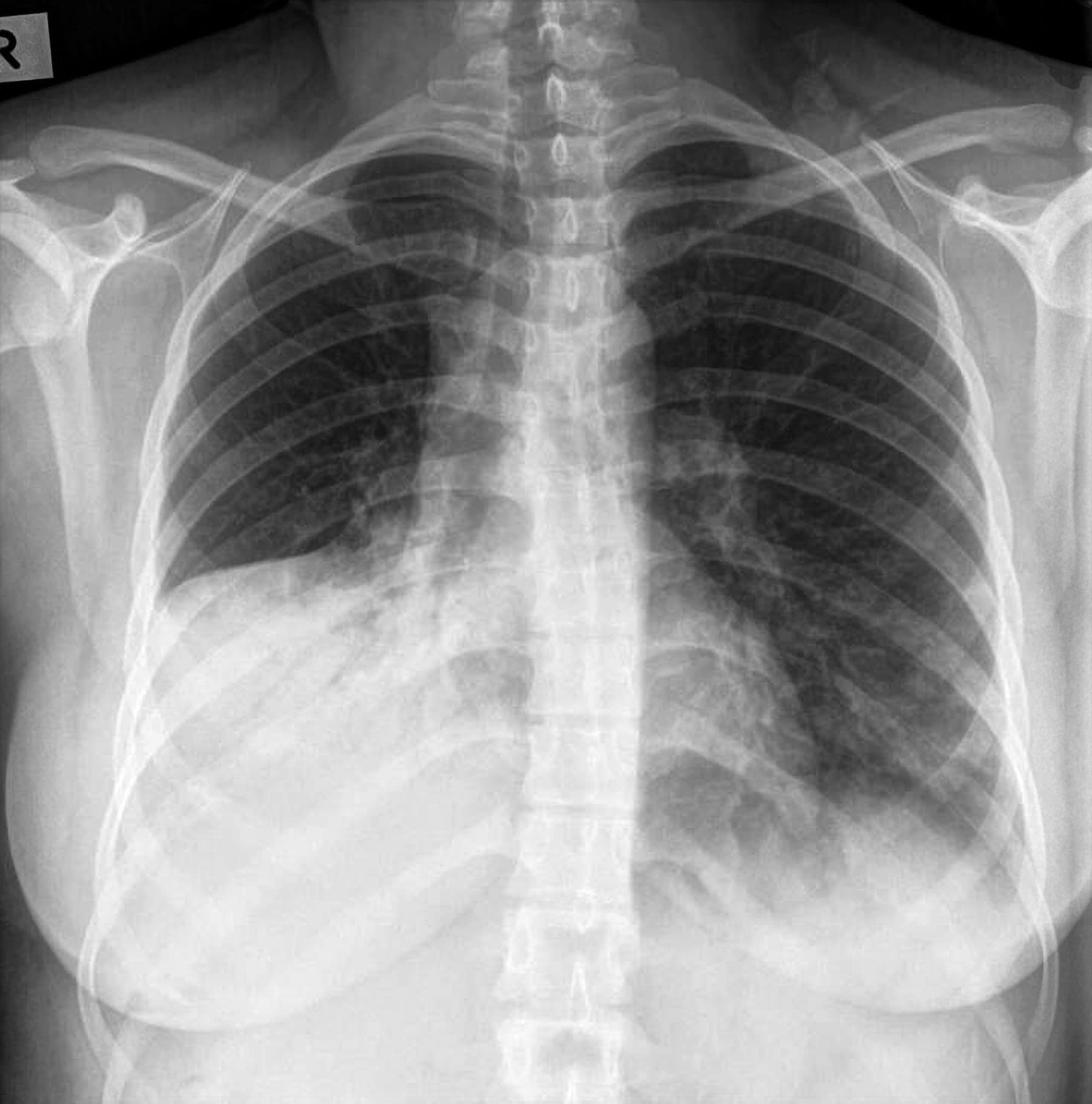
Pneumonia is characterised by exudation and consolidation into the alveoli and in the u k. Erect pa image a and lateral chest x ray image b demonstrates pleural effusion.










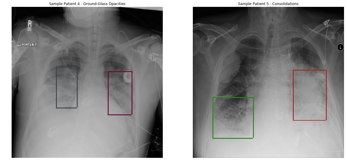
:max_bytes(150000):strip_icc()/covid-19-pneumonia-12-20adbdbe7ee54f7784689c3b1ede2d1c.jpg)
